Showing posts with label neuroscience. Show all posts

Saturday, 15 November 2014

Meet the cells that keep neurons running

by Jonathan Smith

When watching a Formula 1 race, it’s easy to forget that the racing drivers, skilled as they are, don’t work alone. When the car pulls into a pit stop, however, you see a bustling team of mechanics and other experts eagerly rush out to keep the car and driver in top condition. Meanwhile, security staff stand by, blocking public access and keeping a watchful eye on any danger that may present itself. Similarly too, the F1 drivers of the nervous system, neurons, get a lot of attention due to their unique information-processing properties. However, there’s a diverse team of specialised cells that beaver away in the background of the nervous system, carrying out essential tasks analogous to the F1 driver’s pit stop team. Without these plucky little cells, neurons wouldn’t propagate information properly and the nervous system would cease to function. With that in mind then, let’s give some of the most important overlooked cells the limelight for a change, starting with the ‘security guards’ of the mammalian nervous system.

Endothelial cells form the Great Wall of the Brain
As we eat, sleep and move about, our blood ion and sugar levels are constantly in fluctuation. For our neurons that require tightly controlled conditions, exposure to this would be highly detrimental. Thankfully, we can look to endothelial cells, which line the interface between brain tissue and the bloodstream, forming a shield against peripheral influence known as the Blood-Brain Barrier (BBB). By surrounding blood vessels and plugging gaps in the line with protein complexes called Tight Junctions, these endothelial cells and their buddies the pericytes not only cushion brain tissue from fluctuating ion and glucose concentrations but also block passage to many complex molecules, including foreign pathogens.


/static-content/images/480/art%253A10.1007%252Fs10545-013-9608-0/MediaObjects/10545_2013_9608_Fig3_HTML.gif
Figure 1: The endothelial Blood-Brain Barrier around a capillary (red chamber) combines with glial cells such as microglia and astrocytes to form a safe nutrient delivery system to neurons. Pericytes and tight junctions help the endothelial cells to seal the boundary and astrocytes use an end foot process to suck up the nutrients passed along by the barrier. Microglia wait on the side, checking for hazards. Source: SR Yusof and NJ Abbott, from Abbot, 2013, doi: 10.1007/s10545-013-9608-0

Not content to simply act as a wall, these cells also tirelessly shuttle essential ions, sugars and other chemicals into the brain and remove toxins through their own cytoplasm, ensuring that neurons are both protected from the outside world and aptly supplied with the nutrients they require. Furthermore, white blood cells of the immune system - the body’s police force - regularly squeeze through the barrier in order to check for danger. However, this also works against the brain in conditions such as strokes in which the BBB becomes leaky and can’t limit the passage of white blood cells, thus increasing inflammation and exacerbating the problem.

You won’t like microglia when they’re angry
Inside the BBB we encounter the glial cell population. Glial cells - named after the Greek word for ‘glue’ - share many characteristics with neurons but lack the unique structures and functions that specialise neurons for information transmission. Instead, glial cells play a diverse set of roles maintaining the nervous system. Numbering at around 15% of all glial cells, security guard microglia are the subject of intense research because of their incredible versatility. These cells spend the majority of their time simply sitting in the brain tissue, waving their dainty branch-like processes around in a constant search for signs of danger. When they pick up a scent of damage or invasion, however, microglia turn ugly. These sentinels kick off tissue inflammation and undergo an Incredible Hulk-esque transformation into a blob that engulfs and digests the offending party before innocently reverting back to its original state. 


Figure 2: Microglia (green) detects a tissue injury and springs to action. Its extended processes detect signs of damage and trigger the cell’s transformation into a big blob that engulfs the debris. Source: adapted from Nayak et al, 2014, doi: 10.1146/annurev-immunol-032713-120240

With this astounding response to hazards, microglia make impressive enforcers. However, mountains of research have revealed that they can do much more than this. They communicate with cells of the immune system that pop in every so often. In addition, microglia have been found to nurture the growth of neurons in embryonic development and help to prune unwanted cells from the nervous system in developing juveniles. They could even play an important role in synaptic function, making them crucial for normal information processing. However, these eager cells may also work against us in neurodegenerative diseases such as Alzheimer’s Disease and Parkinson’s Disease, believed to involve high inflammation that proves to be neurotoxic in the long run. By better understanding the role these cells play in the pathology, we might able to devise new strategies for treating these conditions.

Insulating the wiring with oligodendrocytes
Mammal neurons are small, thin cells compared to some of the whoppers found in invertebrate animals such as the giant squid. Most neurons output their information through a long process called an axon and, due to electrical resistance inside the cell, neurons with thinner axons propagate information more slowly than those with thicker axons. How then could a nervous system operate with such small neurons? The answer is myelination. This is the process by which specialised glial cells called oligodendrocytes and their peripheral cousins Schwann cells tightly wrap their own fatty membrane - the myelin sheath - around the axons, leaving little gaps of axonal membrane that allow electrical potentials to ‘jump’ significant distances along the axon. This increases possible propagation speeds up to 100 metres per second in humans - perfect for neuronal communication.
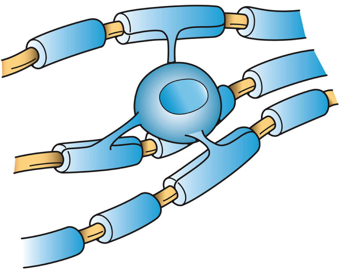

Figure 3: Oligodendrocytes (blue) ensheathe many axons (brown) in myelin to facilitate electrical transmission. Source: Wikipedia

Insulation might not be the only function for these ‘mechanics’ of the nervous system. Recent research indicates that oligodendrocytes can not only insulate axons, but may also directly support the axon’s energy requirements by supplying substrate molecules used in metabolic reactions such as lactate. Further studies indicate that oligodendrocytes promote neuronal survival and axonal growth. The importance of these cells and Schwann cells is further underscored by the fact that the disorder Multiple Sclerosis (MS) arises from demyelination of axons throughout the nervous system. For a multitude of reasons, the immune system attacks myelin and thus deprives neurons of essential support, causing neurodegeneration and, as a consequence of this, ultimately life-threatening paralysis in MS patients. However, strategies are now being trialled that may be able to divert or retrain the immune system and prevent the progression of MS.

Astrocytes - star players in the nervous system
Astrocytes are star-shaped glial cells thanks to their many fine processes, hence the name. Acclaimed as the most abundant type of cell in the human brain, these cells have the chief responsibility of transporting nutrients from blood vessels to nearby neurons by means of a long ‘foot’ process. Astrocytes also oversee chemical synapses - vital junctions at which neurons communicate using neurotransmitters - and each astrocyte can monitor up to a whopping 140,000 synapses! Taking roles analogous to trainers, medics and mechanics in the F1 team, astrocytes are absolutely essential for the survival of the nervous system and by extension, the entire organism.

As can be expected, lots of research gets devoted to unraveling the precise roles that astrocytes play in the nervous system. It’s now clear that these ubiquitous cells encourage the formation and pruning of synapses in the developing brain. In addition, they fine-tune synaptic activity by supplying necessary energy substrates, hoovering up and recycling excess neurotransmitters, prevent seizures by clearing away potassium ions and physically ensheath the synaptic space, reducing spillover of neurotransmitters to nearby cells. Unfortunately, astrocytomas are among the most common cancers in the nervous system and have a relatively high mortality rate. They damage the brain tissue by increasing pressure inside the skull, compete for nutrients and releasing toxic chemicals into the brain, resulting in varied symptoms including headaches, seizures and occasionally personality changes. Though the classic cancer treatments are available such as surgical removal, it’s hard to cut away all of the high-grade tumours due to their rapid infiltration into the brain.

Collective thinking
Aside from the heroes discussed in this piece, there are also tons more subsets of cells that deserve honourable mentions, including parenchymal cells that circulate cerebrospinal fluid around the brain and the somewhat enigmatic NG2 glia, whose precise function still eludes us. While staying mostly in the background, these other cells all have vital functions that serve to ensure that our neurons keep running smoothly. 

Neurons tend to be the main focus of neuroscientific studies. This attention is well deserved considering that they are the substrates of our very thoughts. However, after examining some non-neuronal cells, it’s clear that neurons wouldn’t last a minute without the help of their expert team on hand. By exerting modulatory influence on neuronal development and synaptic activity, it could be argued that the support cells similarly affect our thinking and learning processes, an argument particularly evidenced by complex neurodegenerative diseases involving lots of cell pathologies. With this in mind then, let’s all watch some F1 and spare a thought about the collaborative efforts involved in securing first place!

Sunday, 19 October 2014

Transcranial Direct Current Stimulation: Remoulding the Brain by Duncan Ware


A transient tingling sensation on my scalp, accompanied by an equally fleeting phosphene across my visual field, alerts me to the fact that 2 milliamps of direct current are now passing through my brain, the dorsolateral prefrontal cortex (DLPFC) to be specific. No, I haven’t been denied extradition from a pro-electric chair state, I willingly made myself a component in the circuitry of a technology known as transcranial direct current stimulation (tDCS).

It is widely accepted that everything we do has an effect on the ‘wiring’ of our brains, a fact proposed most succinctly by neuropsychologist Donald Hebb, whose words are forever paraphrased as “neurons that fire together, wire together”. Hebb’s law is now known to rely on long-term potentiation (LTP) and long-term depression (LTD), the enhancement and reduction of synaptic efficacy, respectively. These mechanisms of synaptic plasticity are thought to be the fundamental processes which underlie learning and memory, and perhaps even mood disorders and addiction. It is therefore of little surprise that tDCS, a technology capable of modulating synaptic plasticity, has become subject to a great deal of research in recent years.

TDCS involves the application of electrodes to the scalp above particular regions of the brain, as determined by the Brodmann area map used for electroencephalography (EEG); the regions stimulated dictate the effects of the session. The anode exerts a depolarising influence on the neuronal somata (neuronal cell bodies) of the cortex and hyperpolarises the apical dendrites, whereas the cathode induces hyperpolarisation of the somata and depolarisation of the apical dendrites. Relating this back to synaptic plasticity, the areas affected by the anode become more likely to ‘fire’, meaning their synapses are more prone to LTP, and, conversely, regions of the brain affected by the cathode become less active and are more likely to undergo LTD. This is more or less the extent to our understanding of the mechanism by which the effects of tDCS are manifested.

The clinical applications of an electrical current applied to the scalp have been known for years. As far back as 43 AD, in fact, Roman emperor Claudius’ physician used the shocks of electric eels to abate the pain of headaches! Today it is known that tDCS is capable of ameliorating a multitude of pathological afflictions, from stroke damage to schizophrenia, but also that you and I, as presumably healthy individuals, might derive benefit from the occasional zap.

Attending to the former claim of therapeutic potential in the ill, the montage (electrode placement) with which I am, to use the term most loosely, experimenting today has been found to remediate depression. Some studies have found that just 20 minutes of 2 mA anodal stimulation over the DLPFC to reduce self-reported depression by as much as 10% for every week of daily use. Unfortunately, many of the studies I have come across regarding tDCS are ‘open-label’, science jargon denoting clinical trials in which both the researchers and participants know which subjects are receiving which treatments (in this case, the real treatment or a ‘sham’ control). Unlike its ‘double-blind’ antithesis, open-label studies are plagued by the expectancy effects of both the researcher’s overt enthusiasm, or lack thereof, for the treatment and the subject’s expectations of its outcome. Consequently, one might denounce the aforementioned results to be a direct outcome of the placebo effect. This criticism has been largely dismissed by more recent double-blind trials and studies investigating the relative efficacy of tDCS and established pharmacological therapies such as sertraline (an SSRI antidepressant). Such studies have found tDCS and SSRIs to be of equal efficacy, though a combination of the two was found to be of superior efficacy to either alone, a synergistic pairing I hope will soon be exploited in clinical practice.

For those who refrain from the use of recreational drugs due to their deleterious effects or illegality, perhaps you might consider potentiating your own endogenous substances for a similar effect? I recently came across a most intriguing montage which achieves just that. With the anode attached to the C3 Brodmann area, corresponding to the region of the scalp which lies above the primary motor cortex of the left hemisphere, and the cathode pressed against my upper right arm, effects reminiscent of those one might experience following consumption of a weak opiate such as codeine were elicited almost immediately. As someone who has done much experimenting, in the euphemistic sense, this was a most welcome experience I quickly sought to investigate. A google or two later and I found a publication released last year detailing the analgesic potential of tDCS, an effect they put down to the ยต-opioid system. Opioid receptors are those which transduce the effects of opiates and opioids (substances of similar pharmacological profiles to opiates), like morphine and methadone respectively. But the body possesses its own painkillers, including enkephalins, endorphins and dynorphins to name but a few, and it is these substances whose production is upregulated upon stimulation of the motor cortex. What the paper failed to mention, however, was the euphoric sensations evoked by this montage. Feeling like a character from Huxley’s Brave New World, I amused myself with the idea of becoming a junky without ever having pierced a vein or ‘chased the dragon’.

Another, more frequently studied, area of tDCS research focuses on the technology’s potential as a cognitive enhancer. The phrase is employed with ever increasing frequency as we strive to match our efficiency with the demands of modern life, or perhaps to simply mimic Bradley Cooper’s character in the film ‘Limitless’! Whilst I shan’t delve too deeply into the ethical storm which stalks this phrase, I feel that the practice must be discussed. The majority of research in this area pertains to the augmentation of working memory, the ability to hold information in one’s mind to permit its manipulation and analysis. Such research supports the idea that anodal stimulation of the left prefrontal cortex, a brain region implicated in a variety of executive functions, results in a significant improvement in the working memory of healthy participants. However, some experts admonish users of the trade-off between anodal excitation and cathodal inhibition that is so integral to the device’s mechanism of action. By this, they refer to the fact that whilst you might enhance the activity of one area, with the anode, you will also suppress activity in the area beneath the cathode. Minimising the impact of this trade-off is undoubtedly a task we must prioritise in brain stimulation research, especially given the diffuse nature of tDCS’ influence on the brain, which it seems may extend to subcortical structures.

And so, whilst I must urge you to take caution, should you proceed to plug yourself into the mains (figuratively, a 9 volt battery is sufficient) tDCS is a wonderful medical development which, along with its successors: transcranial magnetic stimulation (TMS) and high-resolution tDCS, I predict we will be seeing much more of in the coming years. 

Thursday, 13 February 2014

Dementia: is a cure around the corner?

by Jonathan Smith

Amidst regular breaking news stories of promising developments in the fight against dementia, we’re still no closer to being able to stop its progression. The company that finds a cure for dementia would not only start a revolution in treatments, but also make huge profits doing so. If this is the case, then why are we still waiting for effective treatments after decades of research into this oppressive disease? 

Prevalence and treatments for dementia

It’s no secret that dementia is a widespread condition. According to the Alzheimer’s Society, the established prevalence of dementia for 70-79 years is 1 in 25. For those over 80, the rate soars to 1 in 6. Considering the colossal cost to healthcare systems worldwide, the potential benefits of finding an effective treatment far outweigh the cost of making it. However, the current treatments available for dementia are minimal. Drugs such as Donepezil, Galantamine and Memantine serve to temporarily reduce the cognitive impairment caused by Alzheimer’s Disease, but do not target the pathology directly. This means that the more advanced cases eventually cease responding to treatment altogether. The brain tissue is just too damaged to salvage by then. What is desperately needed is a treatment that can actually alter the disease itself, resulting in a slower or even halted progression.

It’s not as if pharmaceutical companies haven’t been trying to find disease-modifying treatments of course. Last year the drug Semagecestat failed a massive clinical trial in Alzheimer’s Disease patients. It turned out that patients actually got worse with the drug! In the same year, the catchily-named drug LY2886721 was withdrawn from development due to abnormal liver tests in some trial patients. And failures are costly in the pharmaceutical business. Getting a drug to market can cost millions of pounds and can take over a decade for a company. Thus in the UK at least, industrial research into dementia treatments is getting less funding than ever. This still leaves the question: Why have the drugs failed to work? 

Understanding the neuropathology in dementia

The only simple answer so far is that, being a neurological disease, dementia is an extremely complex puzzle to unravel. Take Alzheimer’s Disease for example. A popular hypothesis of its origin is called the Amyloid Cascade Hypothesis. This suggests that the protein Amyloid-beta starts to get produced abnormally in the brain and clumps together outside cells, gradually causing neurons to malfunction and degenerate, resulting in the symptoms we see in Alzheimer’s Disease. One key piece of evidence for this is that a small proportion of patients get early-onset Alzheimer’s Disease and they all have hereditary mutations causing huge amounts of amyloid-beta production. Not only that, but Down’s Syndrome is also associated with amyloid-beta and guess what? Down’s Syndrome sufferers have a high risk of developing Alzheimer’s Disease. From this evidence, it appeared that a good way to slow Alzheimer’s Disease is by reducing the amyloid-beta production in the brain. As shown by Semagecestat and other failed amyloid-beta-modifying compounds, it turned out to be not that simple.

For one thing, Amyloid-beta is not the only factor to consider in Alzheimer’s Disease. There are many other hypotheses regarding other proteins and cellular systems involved in the symptoms of Alzheimer’s Disease. The result is a rather confusing jumble of different pathologies that neatly overlap with many other types of dementia, such as Vascular Dementia. Additionally, the stage of dementia is a massive factor. Current treatments stop working because there is so much damage to our brain tissue. Perhaps this is also the case with disease-modifying treatments? What if the only way to slow dementia is to tackle it before it causes extensive damage? To investigate this possibility, companies such as Merck are trialling previously failed drugs in patients with early-stage dementia. 

 Amyloid-beta protein
Another consideration is that the best way to trial a treatment early on is to test it in animals like mice and rats. In order to show its effects, we clearly need to give the animal dementia first. To give mice dementia, we first have to insert the mutated genes involved in amyloid-beta production into mice, straight from early-onset patients. As it turns out, it’s really difficult to make mice with Alzheimer’s Disease using genes selected from these patients. Firstly, the vast majority of Alzheimer’s sufferers have no such mutation and secondly, most mouse models of Alzheimer’s Disease show only bits of the condition. Some are cognitively impaired in some tests, others show amyloid-beta clumps, however, none show Alzheimer’s Disease in its entirety. It’s for this reason that many sensational headline cures for dementia found in rodents remain in rodents. It’s just not possible to perfectly translate a complex condition like dementia between rodents and humans. At least, not at the moment.

A cure is around a corner

Despite the recent setbacks in clinical trials, researchers are increasing our knowledge about developing treatments for dementia. Not only that, but steps are being made in improving the diagnosis of Alzheimer’s Disease early on, perhaps at a stage when a difference can be made to the outcome. As animal models of dementia are improved, we can begin to make better translation between animals and human patients. Furthermore, if as much funding for dementia is poured into these efforts as there is for cancer studies, we can increase the speed at which really effective treatments become available. For now, though, the best ways to reduce the risk of dementia are through a balanced diet, regular exercise and regular glasses of red wine!

Friday, 20 December 2013

The Musical Brain

2013 BNA Christmas Symposium overview

by Jonathan Smith

How does the human brain distinguish music from noise? What brain regions are active when we react to music? Do we all share an intrinsic musicality? How do you make a duck into a soul singer?

These important questions were discussed this month in an annual Christmas symposium held by the British Neuroscience Association (BNA). Speakers from all over the UK were invited to present their findings on the special relationship between Homo sapiens and music. These talks were also interspersed with live music, refreshments and humorous ‘Christmas Crackers’ such as the latter question asked above. In this article I summarise the research discussed in this exciting symposium.

Distinguishing music from noise by pattern-detection
It’s rare to be in a completely silent environment, even in university exams! Being able to tell apart rhythm from random noise is very advantageous. This is because we can be alerted to someone’s footsteps for example, which can let us calculate all sorts of useful information such as the proximity, speed and even mood of the walker.

Dr Maria Chait from the University College London (UCL) demonstrated that humans are incredibly sensitive to rhythmic, repeating sounds. This is even the case when our attention is diverted to other tasks - showing that there is continuous, sub-conscious processing that is very effective at detecting rhythms in our auditory inputs. This might go some way to explaining why all human societies feature some form of rhythmic musical tradition, including genres like polyrhythmic African drumming and thumping dance floor beats.

The Beat in society
It’s clear that an important component of most music is a regular pulse, or beat. The beat provides a regular structure on which we can build harmonies, rhythms and melodies. As demonstrated by the audience in a clapping task, humans are very good at detecting the beat of a piece of music and then moving in sync with it - in other words, dancing. Any Youtube video search would also reveal that our fascinating ability starts at an early age. What is happening in the brain when we detect a beat?

In studies by Dr Katie Overy of the University of Edinburgh, participants were tested to see if they could tell if the beat was repeated in patterns of fours, threes or twos, corresponding to 4/4, 3/4 and 2/4 times for musicians. Using fMRI scans to show active brain regions, Dr Katie Overy showed that groups of neurons deep inside the brain called the Basal Ganglia are very active when carrying out this task. The Basal Ganglia are highly connected regions that are really important in both sensory and motor processing, so this might be an interesting link between listening and moving to a beat. Not only this, but diseases involving the Basal Ganglia, such as Parkinson’s Disease, result in impaired beat detection. Perhaps by using music in more therapies we can provide better ways of treating Parkinson’s Disease and other Basal Ganglia disorders.

The emotional response to music
As most would agree, the soundtrack to a film deeply influences how a scene is portrayed. For instance, dissonant melodies convey discomfort and fear whereas smooth, major keys give a sense of calm and peace. At its most extreme, a piece of music can literally make our hairs stand up on end and give us the ‘chills’. This strong emotional response was measured by Dr Alan Watson of Cardiff University.

Dr Alan Watson’s lab used lie detectors to find out when we get the chills from a piece of music. This is due to the fact that lie detectors are very sensitive to changes in autonomic nervous system activity, such as sweating and pulse rate. Since our autonomic nervous system changes in response to strong emotions, the lie detector is a nifty way of showing when we get the chills! Using various imaging studies, the researchers were able to show that the chills are accompanied by a huge release of dopamine in the ‘pleasure’ circuits in the brain. This thus helps to explain why we can react so strongly to music.

Congenital Amusia and musicality
Some individuals are unable to enjoy music. Some, for example, even have trouble distinguishing between Happy Birthday and the National Anthem. These people may suffer from a condition called Congenital Amusia, a disorder of interpreting musical patterns. Yet, studies of these unique individuals may uncover just how innate musicality can be in the human brain. Dr Lauren Stewart from UCL collaborated with the BBC to carry out some of these studies.

Using a test called the Montreal Battery, the researchers found that people with this disorder have difficulty distinguishing musical tones compared with controls. They even have some trouble in detecting changes in speech tones, such as a question or a command. The research got more elaborate. The experimenters designed an artificial nonsense language and asked participants to detect if they heard a particular word in a phrase e.g. Pa-ti-ba. Interestingly, amusics were no different to controls, even when the ‘language‘ was replaced by musical tones! This indicates that amusia-sufferers may not have an absolute deficit in distinguishing pitches, but rather a lower confidence when doing so. This also indicates that a form of musicality is present in all individuals but can be honed by constant practice.

Dementia and music
Most of us are acquainted with someone who is going through the pain of dementia. It’s a very isolating ordeal for all involved and it’s expected to get much more common within the next few decades. Is music a good way of maintaining contact with sufferers who are gradually losing other precious memories?

Dr Jason Warren from UCL began by emphasising the complexity of music as a cognitive function. It’s encoded in many brain regions and evokes strong emotional and associative memories of events of that concert, party etc. All types of dementia have unique patterns of brain region damage. For example, Frontotemporal dementia (FTD) has specific damage in the knowledge-encoding temporal regions and the motor and emotion-encoding frontal regions of the brain. It turns out that FTD patients have selective impairments in identifying scary and angry music. This may prove to be an effective diagnostic tool because music is a much more robust memory than current tests using the memory of faces.


Peter Todd of the Alzheimer’s Society gave a fascinating talk about his experiences. He organises weekly singing groups called Singing for the Brain. The only difference here is that the participants are dementia sufferers at all stages of the disease. While it might not seem easy to pull off a group session with this requirement, the results of these groups are very encouraging. The groups have even performed at festivals and for BBC Radio 4! The aim of the groups is to include everyone at a personal level, no matter what level of dementia they are suffering. One heartwarming example of the good effects of these groups is of one patient who had lost his short-term memory. He couldn’t even remember that he had been in a singing group for the last hour! However, after every session, it was clear from his posture and manner that he was very upbeat from singing with the group, despite not being able to remember why! Examples like this emphasise the importance of music in social bonding for potentially lonely individuals going through dementia.

Wrap-up
It’s clear that music has been an integral part of human history. This shown by the presence of music in every human culture on Earth and the sheer amount of processing power devoted to music in our brains. The brain is a pattern-seeking machine and it has progressed from interpreting primitive vocalisations in forests to sophisticated music forms. Our emotional connection to music and musicality is preserved to a certain extent in everyone. It also proves to be an effective tool for identifying dementia symptoms and also encourages social inclusion for dementia sufferers.

Oh, and if anyone was curious about how you turn a duck into a soul singer, the answer is: Put it in the microwave until its Bill Withers.