Showing posts with label medicine. Show all posts

Saturday, 15 November 2014

Meet the cells that keep neurons running

by Jonathan Smith

When watching a Formula 1 race, it’s easy to forget that the racing drivers, skilled as they are, don’t work alone. When the car pulls into a pit stop, however, you see a bustling team of mechanics and other experts eagerly rush out to keep the car and driver in top condition. Meanwhile, security staff stand by, blocking public access and keeping a watchful eye on any danger that may present itself. Similarly too, the F1 drivers of the nervous system, neurons, get a lot of attention due to their unique information-processing properties. However, there’s a diverse team of specialised cells that beaver away in the background of the nervous system, carrying out essential tasks analogous to the F1 driver’s pit stop team. Without these plucky little cells, neurons wouldn’t propagate information properly and the nervous system would cease to function. With that in mind then, let’s give some of the most important overlooked cells the limelight for a change, starting with the ‘security guards’ of the mammalian nervous system.

Endothelial cells form the Great Wall of the Brain
As we eat, sleep and move about, our blood ion and sugar levels are constantly in fluctuation. For our neurons that require tightly controlled conditions, exposure to this would be highly detrimental. Thankfully, we can look to endothelial cells, which line the interface between brain tissue and the bloodstream, forming a shield against peripheral influence known as the Blood-Brain Barrier (BBB). By surrounding blood vessels and plugging gaps in the line with protein complexes called Tight Junctions, these endothelial cells and their buddies the pericytes not only cushion brain tissue from fluctuating ion and glucose concentrations but also block passage to many complex molecules, including foreign pathogens.


/static-content/images/480/art%253A10.1007%252Fs10545-013-9608-0/MediaObjects/10545_2013_9608_Fig3_HTML.gif
Figure 1: The endothelial Blood-Brain Barrier around a capillary (red chamber) combines with glial cells such as microglia and astrocytes to form a safe nutrient delivery system to neurons. Pericytes and tight junctions help the endothelial cells to seal the boundary and astrocytes use an end foot process to suck up the nutrients passed along by the barrier. Microglia wait on the side, checking for hazards. Source: SR Yusof and NJ Abbott, from Abbot, 2013, doi: 10.1007/s10545-013-9608-0

Not content to simply act as a wall, these cells also tirelessly shuttle essential ions, sugars and other chemicals into the brain and remove toxins through their own cytoplasm, ensuring that neurons are both protected from the outside world and aptly supplied with the nutrients they require. Furthermore, white blood cells of the immune system - the body’s police force - regularly squeeze through the barrier in order to check for danger. However, this also works against the brain in conditions such as strokes in which the BBB becomes leaky and can’t limit the passage of white blood cells, thus increasing inflammation and exacerbating the problem.

You won’t like microglia when they’re angry
Inside the BBB we encounter the glial cell population. Glial cells - named after the Greek word for ‘glue’ - share many characteristics with neurons but lack the unique structures and functions that specialise neurons for information transmission. Instead, glial cells play a diverse set of roles maintaining the nervous system. Numbering at around 15% of all glial cells, security guard microglia are the subject of intense research because of their incredible versatility. These cells spend the majority of their time simply sitting in the brain tissue, waving their dainty branch-like processes around in a constant search for signs of danger. When they pick up a scent of damage or invasion, however, microglia turn ugly. These sentinels kick off tissue inflammation and undergo an Incredible Hulk-esque transformation into a blob that engulfs and digests the offending party before innocently reverting back to its original state. 


Figure 2: Microglia (green) detects a tissue injury and springs to action. Its extended processes detect signs of damage and trigger the cell’s transformation into a big blob that engulfs the debris. Source: adapted from Nayak et al, 2014, doi: 10.1146/annurev-immunol-032713-120240

With this astounding response to hazards, microglia make impressive enforcers. However, mountains of research have revealed that they can do much more than this. They communicate with cells of the immune system that pop in every so often. In addition, microglia have been found to nurture the growth of neurons in embryonic development and help to prune unwanted cells from the nervous system in developing juveniles. They could even play an important role in synaptic function, making them crucial for normal information processing. However, these eager cells may also work against us in neurodegenerative diseases such as Alzheimer’s Disease and Parkinson’s Disease, believed to involve high inflammation that proves to be neurotoxic in the long run. By better understanding the role these cells play in the pathology, we might able to devise new strategies for treating these conditions.

Insulating the wiring with oligodendrocytes
Mammal neurons are small, thin cells compared to some of the whoppers found in invertebrate animals such as the giant squid. Most neurons output their information through a long process called an axon and, due to electrical resistance inside the cell, neurons with thinner axons propagate information more slowly than those with thicker axons. How then could a nervous system operate with such small neurons? The answer is myelination. This is the process by which specialised glial cells called oligodendrocytes and their peripheral cousins Schwann cells tightly wrap their own fatty membrane - the myelin sheath - around the axons, leaving little gaps of axonal membrane that allow electrical potentials to ‘jump’ significant distances along the axon. This increases possible propagation speeds up to 100 metres per second in humans - perfect for neuronal communication.
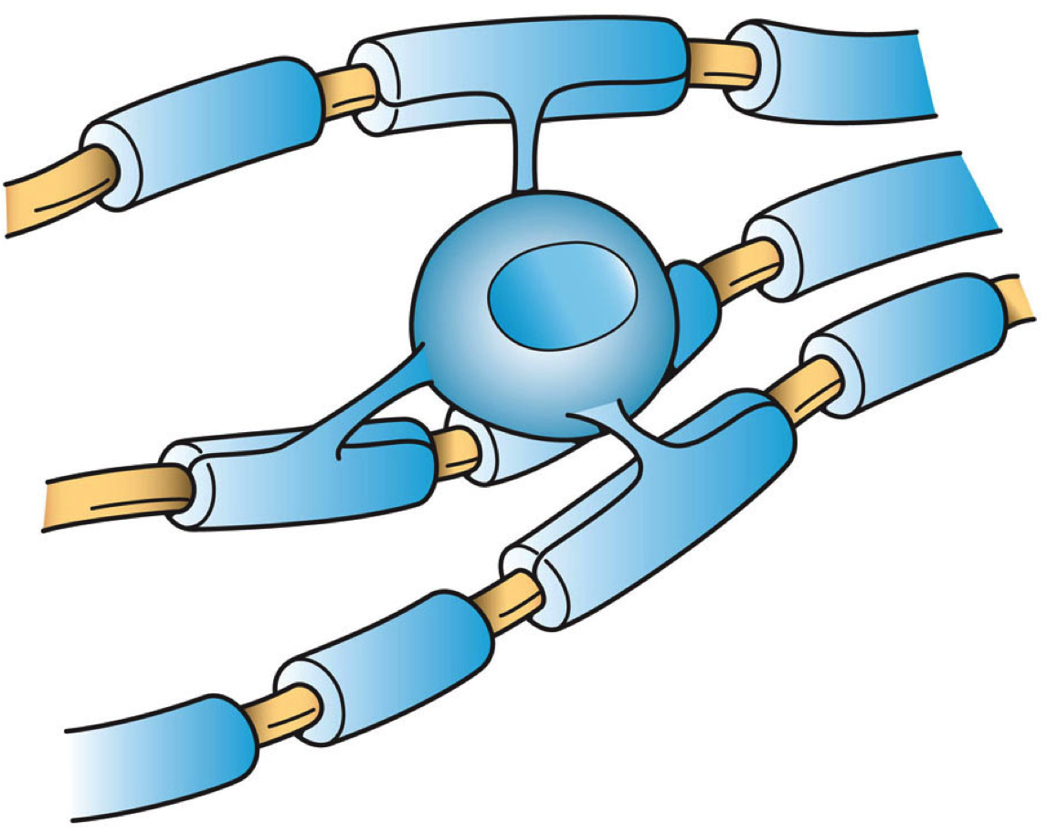

Figure 3: Oligodendrocytes (blue) ensheathe many axons (brown) in myelin to facilitate electrical transmission. Source: Wikipedia

Insulation might not be the only function for these ‘mechanics’ of the nervous system. Recent research indicates that oligodendrocytes can not only insulate axons, but may also directly support the axon’s energy requirements by supplying substrate molecules used in metabolic reactions such as lactate. Further studies indicate that oligodendrocytes promote neuronal survival and axonal growth. The importance of these cells and Schwann cells is further underscored by the fact that the disorder Multiple Sclerosis (MS) arises from demyelination of axons throughout the nervous system. For a multitude of reasons, the immune system attacks myelin and thus deprives neurons of essential support, causing neurodegeneration and, as a consequence of this, ultimately life-threatening paralysis in MS patients. However, strategies are now being trialled that may be able to divert or retrain the immune system and prevent the progression of MS.

Astrocytes - star players in the nervous system
Astrocytes are star-shaped glial cells thanks to their many fine processes, hence the name. Acclaimed as the most abundant type of cell in the human brain, these cells have the chief responsibility of transporting nutrients from blood vessels to nearby neurons by means of a long ‘foot’ process. Astrocytes also oversee chemical synapses - vital junctions at which neurons communicate using neurotransmitters - and each astrocyte can monitor up to a whopping 140,000 synapses! Taking roles analogous to trainers, medics and mechanics in the F1 team, astrocytes are absolutely essential for the survival of the nervous system and by extension, the entire organism.

As can be expected, lots of research gets devoted to unraveling the precise roles that astrocytes play in the nervous system. It’s now clear that these ubiquitous cells encourage the formation and pruning of synapses in the developing brain. In addition, they fine-tune synaptic activity by supplying necessary energy substrates, hoovering up and recycling excess neurotransmitters, prevent seizures by clearing away potassium ions and physically ensheath the synaptic space, reducing spillover of neurotransmitters to nearby cells. Unfortunately, astrocytomas are among the most common cancers in the nervous system and have a relatively high mortality rate. They damage the brain tissue by increasing pressure inside the skull, compete for nutrients and releasing toxic chemicals into the brain, resulting in varied symptoms including headaches, seizures and occasionally personality changes. Though the classic cancer treatments are available such as surgical removal, it’s hard to cut away all of the high-grade tumours due to their rapid infiltration into the brain.

Collective thinking
Aside from the heroes discussed in this piece, there are also tons more subsets of cells that deserve honourable mentions, including parenchymal cells that circulate cerebrospinal fluid around the brain and the somewhat enigmatic NG2 glia, whose precise function still eludes us. While staying mostly in the background, these other cells all have vital functions that serve to ensure that our neurons keep running smoothly. 

Neurons tend to be the main focus of neuroscientific studies. This attention is well deserved considering that they are the substrates of our very thoughts. However, after examining some non-neuronal cells, it’s clear that neurons wouldn’t last a minute without the help of their expert team on hand. By exerting modulatory influence on neuronal development and synaptic activity, it could be argued that the support cells similarly affect our thinking and learning processes, an argument particularly evidenced by complex neurodegenerative diseases involving lots of cell pathologies. With this in mind then, let’s all watch some F1 and spare a thought about the collaborative efforts involved in securing first place!

Saturday, 10 May 2014

Transition in Pharma

What needs to be prescribed to an industry in distress?

by Toby Benham


Recent large scale closures of R&D sites in the UK from pharma giants Pfizer, Merck, GSK and now Novartis has led to the nationwide desolation of the pharmaceutical industry. With cuts extending around the world, and several big challenges ahead, it appears the industry is heading into a time of transition. To emerge through this transition stronger it is important for pharmaceutical companies to collaborate, working together for the collective good of the field.

Difficult times
The dominant business model adopted in recent times by pharmaceutical companies involved investing heavily into promising drug candidates, attempting to create the next big blockbuster. For associated with these iconic blockbusters are fame and fortune. Drugs such as Lipitor and Plavix have allowed their respective companies to thrive previously. However, the industry has been looming over the edge of the “patent cliff” (when many current blockbuster patents expire) for several years and now companies are lining up for the plunge. It means that these drugs can be manufactured and sold by any generics company at the detriment of the inventor company’s profits. This strategy relies on new blockbusters to come through the system but current pipelines appear relatively fruitless. 

Developing new drugs is an expensive business. Forbes estimates that it now costs approximately $5 billion per new drug created; this is not a sustainable figure. Costs spiral during the 15 years that contribute to getting a drug to market. The drug discovery, optimisation, clinical trials, patent protection and marketing involved are all long expensive processes. However, the main reason that the figure is so high is due to the unseen added cost of research into unsuccessful drug projects. Thus, there could not be a worse time for worldwide scandals to be breaking out in the news, smearing the image of pharma. Just last year, both GSK and Novartis were alleged to have bribed doctors and healthcare officials in China. There are also questions over the safety of some drugs already on the market.  GSK’s “Avandia” for diabetes treatment has been under intense scrutiny for several years now with restrictions in the US only lifted recently. With so many hurdles in the development process - ranging from toxicity to manufacturing - high risk, high reward projects may now be considered just that bit too risky. 

The future
Most importantly, big pharma need to ditch their profit alone method and support one another for their collective benefit. In 2013, data analytics company SAS announced the creation of a globally accessible but private bank of data for pharmaceutical companies to pool clinical trial data. GSK have been the first to share. Perry Nisen, the GSK senior vice president for science and innovation, announced that, “in sharing our data with researchers across the world, we hope to further scientific research and increase understanding about our medicines.”  This exemplary collaborative model will allow companies to improve efficiency and enhance the decision making progress which is so crucial in pushing forward drug candidates. Working on projects across companies should also be encouraged with the chance to explore new opportunities, widen portfolios and spread risk. GSK and Novartis recently announced an asset swapping deal, but this could go even further.


In addition, the big pharmaceutical companies can collaborate with the smaller businesses to flourish from symbiotic relationships. Companies such as Aurigene offer cost effective outsourcing of R&D in their respective areas of expertise, creating what Aurigene describe as a “win-win partnership” that accelerates discovery. The opportunities are not limited to industry with many experts in academia to link up with. Sanofi-Aventis and Pfizer have already created strong partnerships for drug development with Harvard University and UCFS respectively. Back in the UK, Astra Zeneca is building a new headquarters located in Cambridge with the intent to partner with Cambridge University and local hospitals.  By sharing scientific talent and resources, the drug development process gains extra quality and creativity from fresh perspectives. Diversity and partnerships lead to innovation which is essential to feeding hungry company pipelines. A wider communication with regulators would also be invaluable. Hopefully this could put an end to public scandals and improve the clinical trial process. 

Change is required to replace the current unsustainable business model in the pharmaceutical industry. With the right partnerships, a new streamlined, cost effective and innovative R&D system is possible. This will reduce the price of creating a drug by increasing productivity whilst simultaneously cutting expenditures. Through sharing scientific talent, resources and knowledge it is possible for the industry to return from the drop of the patent cliff to emerge stronger by optimising the potential of collaboration. Pharmaceutical companies should consider working in unison for the common goal and share the rewards. This is important not just for the companies concerned but for the patients that benefit as a result.

Thursday, 13 February 2014

Dementia: is a cure around the corner?

by Jonathan Smith

Amidst regular breaking news stories of promising developments in the fight against dementia, we’re still no closer to being able to stop its progression. The company that finds a cure for dementia would not only start a revolution in treatments, but also make huge profits doing so. If this is the case, then why are we still waiting for effective treatments after decades of research into this oppressive disease? 

Prevalence and treatments for dementia

It’s no secret that dementia is a widespread condition. According to the Alzheimer’s Society, the established prevalence of dementia for 70-79 years is 1 in 25. For those over 80, the rate soars to 1 in 6. Considering the colossal cost to healthcare systems worldwide, the potential benefits of finding an effective treatment far outweigh the cost of making it. However, the current treatments available for dementia are minimal. Drugs such as Donepezil, Galantamine and Memantine serve to temporarily reduce the cognitive impairment caused by Alzheimer’s Disease, but do not target the pathology directly. This means that the more advanced cases eventually cease responding to treatment altogether. The brain tissue is just too damaged to salvage by then. What is desperately needed is a treatment that can actually alter the disease itself, resulting in a slower or even halted progression.

It’s not as if pharmaceutical companies haven’t been trying to find disease-modifying treatments of course. Last year the drug Semagecestat failed a massive clinical trial in Alzheimer’s Disease patients. It turned out that patients actually got worse with the drug! In the same year, the catchily-named drug LY2886721 was withdrawn from development due to abnormal liver tests in some trial patients. And failures are costly in the pharmaceutical business. Getting a drug to market can cost millions of pounds and can take over a decade for a company. Thus in the UK at least, industrial research into dementia treatments is getting less funding than ever. This still leaves the question: Why have the drugs failed to work? 

Understanding the neuropathology in dementia

The only simple answer so far is that, being a neurological disease, dementia is an extremely complex puzzle to unravel. Take Alzheimer’s Disease for example. A popular hypothesis of its origin is called the Amyloid Cascade Hypothesis. This suggests that the protein Amyloid-beta starts to get produced abnormally in the brain and clumps together outside cells, gradually causing neurons to malfunction and degenerate, resulting in the symptoms we see in Alzheimer’s Disease. One key piece of evidence for this is that a small proportion of patients get early-onset Alzheimer’s Disease and they all have hereditary mutations causing huge amounts of amyloid-beta production. Not only that, but Down’s Syndrome is also associated with amyloid-beta and guess what? Down’s Syndrome sufferers have a high risk of developing Alzheimer’s Disease. From this evidence, it appeared that a good way to slow Alzheimer’s Disease is by reducing the amyloid-beta production in the brain. As shown by Semagecestat and other failed amyloid-beta-modifying compounds, it turned out to be not that simple.

For one thing, Amyloid-beta is not the only factor to consider in Alzheimer’s Disease. There are many other hypotheses regarding other proteins and cellular systems involved in the symptoms of Alzheimer’s Disease. The result is a rather confusing jumble of different pathologies that neatly overlap with many other types of dementia, such as Vascular Dementia. Additionally, the stage of dementia is a massive factor. Current treatments stop working because there is so much damage to our brain tissue. Perhaps this is also the case with disease-modifying treatments? What if the only way to slow dementia is to tackle it before it causes extensive damage? To investigate this possibility, companies such as Merck are trialling previously failed drugs in patients with early-stage dementia. 

 Amyloid-beta protein
Another consideration is that the best way to trial a treatment early on is to test it in animals like mice and rats. In order to show its effects, we clearly need to give the animal dementia first. To give mice dementia, we first have to insert the mutated genes involved in amyloid-beta production into mice, straight from early-onset patients. As it turns out, it’s really difficult to make mice with Alzheimer’s Disease using genes selected from these patients. Firstly, the vast majority of Alzheimer’s sufferers have no such mutation and secondly, most mouse models of Alzheimer’s Disease show only bits of the condition. Some are cognitively impaired in some tests, others show amyloid-beta clumps, however, none show Alzheimer’s Disease in its entirety. It’s for this reason that many sensational headline cures for dementia found in rodents remain in rodents. It’s just not possible to perfectly translate a complex condition like dementia between rodents and humans. At least, not at the moment.

A cure is around a corner

Despite the recent setbacks in clinical trials, researchers are increasing our knowledge about developing treatments for dementia. Not only that, but steps are being made in improving the diagnosis of Alzheimer’s Disease early on, perhaps at a stage when a difference can be made to the outcome. As animal models of dementia are improved, we can begin to make better translation between animals and human patients. Furthermore, if as much funding for dementia is poured into these efforts as there is for cancer studies, we can increase the speed at which really effective treatments become available. For now, though, the best ways to reduce the risk of dementia are through a balanced diet, regular exercise and regular glasses of red wine!

Monday, 10 February 2014

Is it safe to reuse plastic bottles?

by Rachel Argo

Typically, I will buy a bottle of water and re-use it until I loose it. We all know that re-using a plastic bottle is good for both the environment and our pockets, but by doing this are we increasing our risk of exposure to ‘dangerous’ chemicals in the plastics? Recently bottles have been designed and marketed as being ‘BPA free’. But what is BPA and why have we never heard of it before?

Maybe you have seen strange shaped bottles with coloured filters appearing in people’s handbags, at the gym or on the high street? ‘Bobble’ bottles are an example of these BPA free plastic products. They are made from recycled materials, are recyclable and contain replaceable filters that claim to remove impurities in the water and improve the taste. Bisphenol A (BPA) is a synthetic organic molecule that is used to make certain plastics such as ‘polycarbonate’. Polycarbonate is commonly used to make household items like drinks bottles and tupperware. BPA is also found in epoxy resins that are used to line drinks cans and tins. We come into contact with plastic all the time and admittedly it would be very difficult to exclude from our day-to-day lives. Until recently no one questioned the effect of this plastic heavy lifestyle on our health, so should we stop re-using that water bottle and buy a BPA free one?

Studies have suggested that BPA possesses the ability to pass from the containers to the food or beverages inside. The concern around this possible seeping stems from the chemical’s ability to act as a mimic of the hormone oestradiol and therefore have potential to interrupt hormone patterns and signaling pathways. Animal studies in rats and mice have linked BPA exposure to a range of health problems such as obesity, fertility impairment, respiratory disorders and inflammation, however there is little or no research into the effects on humans and no single study that conclusively proves that BPA is the cause of these diseases. 

A review in 2006 by the European Food Safety Authority (EFSA), concluded that the Tolerable Daily Intake (TDI) of BPA was 0.05 mg/kg body weight/day. This value is an estimation of the amount of BPA that can be ingested per day per kg of body weight daily, over a lifetime without significant risk to health. BPA is licensed by the EU for use in food contact materials, however a directive in January 2011 prevented the use of BPA containing plastics in the manufacture of baby bottles. The most recent review of the molecule’s safety (July 2013) provisionally suggests that diet is the main source of exposure, that the estimation of this exposure is much lower than EFSAs previous estimations and well below the suggested TDIs. 

Due to the lack of conclusive evidence of BPA safety in humans and changing opinion on TDI values, it is hard to decide if it is worth investing in a BPA free bottle. I initially thought that this was a trendy case of false science, but on closer inspection it seems there may be some truth in their safety claims even if we have not established the extent of it.

Friday, 20 December 2013

The Musical Brain

2013 BNA Christmas Symposium overview

by Jonathan Smith

How does the human brain distinguish music from noise? What brain regions are active when we react to music? Do we all share an intrinsic musicality? How do you make a duck into a soul singer?

These important questions were discussed this month in an annual Christmas symposium held by the British Neuroscience Association (BNA). Speakers from all over the UK were invited to present their findings on the special relationship between Homo sapiens and music. These talks were also interspersed with live music, refreshments and humorous ‘Christmas Crackers’ such as the latter question asked above. In this article I summarise the research discussed in this exciting symposium.

Distinguishing music from noise by pattern-detection
It’s rare to be in a completely silent environment, even in university exams! Being able to tell apart rhythm from random noise is very advantageous. This is because we can be alerted to someone’s footsteps for example, which can let us calculate all sorts of useful information such as the proximity, speed and even mood of the walker.

Dr Maria Chait from the University College London (UCL) demonstrated that humans are incredibly sensitive to rhythmic, repeating sounds. This is even the case when our attention is diverted to other tasks - showing that there is continuous, sub-conscious processing that is very effective at detecting rhythms in our auditory inputs. This might go some way to explaining why all human societies feature some form of rhythmic musical tradition, including genres like polyrhythmic African drumming and thumping dance floor beats.

The Beat in society
It’s clear that an important component of most music is a regular pulse, or beat. The beat provides a regular structure on which we can build harmonies, rhythms and melodies. As demonstrated by the audience in a clapping task, humans are very good at detecting the beat of a piece of music and then moving in sync with it - in other words, dancing. Any Youtube video search would also reveal that our fascinating ability starts at an early age. What is happening in the brain when we detect a beat?

In studies by Dr Katie Overy of the University of Edinburgh, participants were tested to see if they could tell if the beat was repeated in patterns of fours, threes or twos, corresponding to 4/4, 3/4 and 2/4 times for musicians. Using fMRI scans to show active brain regions, Dr Katie Overy showed that groups of neurons deep inside the brain called the Basal Ganglia are very active when carrying out this task. The Basal Ganglia are highly connected regions that are really important in both sensory and motor processing, so this might be an interesting link between listening and moving to a beat. Not only this, but diseases involving the Basal Ganglia, such as Parkinson’s Disease, result in impaired beat detection. Perhaps by using music in more therapies we can provide better ways of treating Parkinson’s Disease and other Basal Ganglia disorders.

The emotional response to music
As most would agree, the soundtrack to a film deeply influences how a scene is portrayed. For instance, dissonant melodies convey discomfort and fear whereas smooth, major keys give a sense of calm and peace. At its most extreme, a piece of music can literally make our hairs stand up on end and give us the ‘chills’. This strong emotional response was measured by Dr Alan Watson of Cardiff University.

Dr Alan Watson’s lab used lie detectors to find out when we get the chills from a piece of music. This is due to the fact that lie detectors are very sensitive to changes in autonomic nervous system activity, such as sweating and pulse rate. Since our autonomic nervous system changes in response to strong emotions, the lie detector is a nifty way of showing when we get the chills! Using various imaging studies, the researchers were able to show that the chills are accompanied by a huge release of dopamine in the ‘pleasure’ circuits in the brain. This thus helps to explain why we can react so strongly to music.

Congenital Amusia and musicality
Some individuals are unable to enjoy music. Some, for example, even have trouble distinguishing between Happy Birthday and the National Anthem. These people may suffer from a condition called Congenital Amusia, a disorder of interpreting musical patterns. Yet, studies of these unique individuals may uncover just how innate musicality can be in the human brain. Dr Lauren Stewart from UCL collaborated with the BBC to carry out some of these studies.

Using a test called the Montreal Battery, the researchers found that people with this disorder have difficulty distinguishing musical tones compared with controls. They even have some trouble in detecting changes in speech tones, such as a question or a command. The research got more elaborate. The experimenters designed an artificial nonsense language and asked participants to detect if they heard a particular word in a phrase e.g. Pa-ti-ba. Interestingly, amusics were no different to controls, even when the ‘language‘ was replaced by musical tones! This indicates that amusia-sufferers may not have an absolute deficit in distinguishing pitches, but rather a lower confidence when doing so. This also indicates that a form of musicality is present in all individuals but can be honed by constant practice.

Dementia and music
Most of us are acquainted with someone who is going through the pain of dementia. It’s a very isolating ordeal for all involved and it’s expected to get much more common within the next few decades. Is music a good way of maintaining contact with sufferers who are gradually losing other precious memories?

Dr Jason Warren from UCL began by emphasising the complexity of music as a cognitive function. It’s encoded in many brain regions and evokes strong emotional and associative memories of events of that concert, party etc. All types of dementia have unique patterns of brain region damage. For example, Frontotemporal dementia (FTD) has specific damage in the knowledge-encoding temporal regions and the motor and emotion-encoding frontal regions of the brain. It turns out that FTD patients have selective impairments in identifying scary and angry music. This may prove to be an effective diagnostic tool because music is a much more robust memory than current tests using the memory of faces.


Peter Todd of the Alzheimer’s Society gave a fascinating talk about his experiences. He organises weekly singing groups called Singing for the Brain. The only difference here is that the participants are dementia sufferers at all stages of the disease. While it might not seem easy to pull off a group session with this requirement, the results of these groups are very encouraging. The groups have even performed at festivals and for BBC Radio 4! The aim of the groups is to include everyone at a personal level, no matter what level of dementia they are suffering. One heartwarming example of the good effects of these groups is of one patient who had lost his short-term memory. He couldn’t even remember that he had been in a singing group for the last hour! However, after every session, it was clear from his posture and manner that he was very upbeat from singing with the group, despite not being able to remember why! Examples like this emphasise the importance of music in social bonding for potentially lonely individuals going through dementia.

Wrap-up
It’s clear that music has been an integral part of human history. This shown by the presence of music in every human culture on Earth and the sheer amount of processing power devoted to music in our brains. The brain is a pattern-seeking machine and it has progressed from interpreting primitive vocalisations in forests to sophisticated music forms. Our emotional connection to music and musicality is preserved to a certain extent in everyone. It also proves to be an effective tool for identifying dementia symptoms and also encourages social inclusion for dementia sufferers.

Oh, and if anyone was curious about how you turn a duck into a soul singer, the answer is: Put it in the microwave until its Bill Withers.

Monday, 11 November 2013

The science of mind reading

by Tom Ridler


The idea of someone being able to tell exactly what we are thinking is no doubt a scary one, but don’t worry, we’re not there yet. This said, the electroencephalogram, or EEG has been used for some time to measure brain activity in human patients and there is a great deal of information to be obtained from all those wiggly lines.

How does EEG work? Our brains are made up of billions of neurons, communicating with each other all of the time. Brain cells “talk” through synapses, creating tiny electrical signals. With so many cells in the brain, this produces masses of electrical activity and it can be measured by placing sensors on the surface of the skull.  This is usually done with the familiar EEG cap, containing a great number of sensors, meaning that different areas of the brain can be measured simultaneously.

What can you see? What we find when we record this brain activity is that the signal within the brain oscillates in wave-like manner. These brain waves may originally seem confusing and random, but analysis has shown that they can be isolated into discrete frequency bands. You can think of the brain like an orchestra, with all the individual instruments creating different sounds that all come together into one complex piece of music.

What does it all mean? These common frequencies may represent differences in brain states. For example, when you are in deep sleep slow oscillations are seen (called delta waves) or during high levels of concentration fast waves (such as beta or gamma oscillations) may occur, signifying intense thought processing. How about some meditation? Well you won’t be doing that without plenty of alpha waves, associated with relaxation and reflection.  

How can it be used? EEG can be used in a great number of ways. We can diagnose some conditions such as epilepsy by recognising seizure activity. There is also potential to help suffers of locked in syndrome (a condition where sufferers, while totally conscious, cannot move or communicate). On a lighter note, many people have been working on ways in which we can control objects with our minds. Just imagine, a brain-machine interface would be able to control a robot, unlock a car or turn on a home appliance just through the power of thought. This isn’t so far of, your own portable (and affordable) EEG machines are available to buy, allowing you to play games and even control the plot of a film through changes in your brain waves. 

Thursday, 24 October 2013

Transplanting Memories?

by Rhema Anderews

George Bernard Shaw once said, “All great truths begin as blasphemies.” In the realm of heart transplantation technology, none has posed greater uproar than the controversial concept of cellular memory.

Cellular memory is the notion that the brain is not the only organ capable of storing memories. In fact, all living cells possess “memory”. Evidence for this has been found predominantly in heart transplant patients. Studies on cellular memory from transplant patients are often conducted by scientists with the aid of the hospital system which forbids the recipient to know or communicate with the donor’s family with most cases without the mention of names.

On May 29, 1988, Claire Sylvia received both the heart and lung of an 18-year-old man killed in a motorcycle accident. After the surgery, Sylvia claimed an intense craving for beer, chicken nuggets and green peppers, all of which she never liked before. She began to assume a masculine walk (peculiar for the dancer), started swearing in conversations, and for no apparent reason took up motorcycle riding at dangerous speeds, which was totally out of character. Sylvia even started having recurring dreams of a mysterious man. In her book entitled “A Change of Heart”, she recounted a dream where she kissed a boy thought to be named Tim L. and inhaled him into her. Upon meeting the “family of her heart” as she put it, Sylvia learned the name of her donor was in fact Tim L., and all of the changes she experienced closely mirrored that of Tim L. who strangely at the point of death had chicken nuggets in his pockets. Sylvia’s story quickly captured media attention and soon after, many other transplant recipients came forward with similar testimonies.

The most striking example is that of an eight-year-old girl who received the heart of a ten year-old-girl. Post-surgery, she was consistently plagued with distressing dreams of an attacker and a girl being murdered. Her nightmares proved so vivid that even her psychiatrist believed them to be genuine memories. As it turns out, the donor was a murder victim and as a result of the recipient’s violent recurring dreams, she was able to describe the horrifying incident and the murderer to such great detail that the police eventually apprehended, arrested and convicted the killer.

Ongoing research has shown that neuropeptides and receptors previously known to exist exclusively in the brain have been discovered in places throughout the body, especially in major organs such as the heart. These neuropeptides are a means for the brain to communicate with other organs and for these organs to send feedback to the brain. However, little is known about whether these neuropeptides can store memory; due to the amount of peptides in the heart, there seems to be a strong correlation between the two. But if this were the case, then why don’t all patients go through this experience?

There is no solid evidence that the reports are nothing more than coincidence and fantasy. Even so, the stories are intriguing and we should expect some serious investigation into the matter in the near future. Until then let’s keep an open heart.

Friday, 11 October 2013

Lip locking, tonsil tennis and infectious mononucleosis: stop smooching?

by Chloe Palmer 

Infectious mononucleosis (IM), more commonly known as glandular fever or ‘the kissing disease’ is an extremely contagious viral disease predominantly caused by the Epstein-Barr Virus (EBV).  Sprunt and Evans in the Bulletin of the Johns Hopkins Hospital first described IM in 1920, however the association between this and EBV was not defined until the late 1960’s. EBV is a type of Herpes virus (Human Herpes Virus-4) containing double-stranded DNA, an icosahedral capsid and a glycoprotein-containing envelope. IM is spread via saliva, and just like all Herpes viruses they become a life companion…or not. The virus has an initial incubation period so an individual may be unaware that they are infected until 4-8 weeks later.  By this point, however, you can be sure that your oropharangeal epithelial cells and B-lymphocytes have taken a massive beating.

IM primarily and most commonly affects teenagers and young adults between the ages of 15-25, probably because school socials and university are saliva-swapping playgrounds (unless you do a science degree in which case you probably don’t get out as much). Initially this sub-clinical infection is asymptomatic but as it progresses characteristic symptoms include a sore throat, fatigue, prolonged malaise, swollen lymph nodes, vomiting, muscle and headaches high fever, a lack of energy and a loss of appetite. Diagnosis may be clear from the symptoms but this can be confirmed by a blood test.

Although both mortality and morbidity rates are low, according to the World Health Organization (WHO) serologic tests have shown that approximately 95% of adults worldwide have been infected with EBV.

There is currently no available treatment for this viral infection, however an EBV vaccine is currently in clinical trials, targeting its envelope glycoproteins (gp350/220). Not only will this be a break-through in preventing IM but also decrease the risk of other associated EBV infections such as Burkitt’s lymphoma and nasopharangeal carcinoma. For now pain relief pills...yes, the same ibuprofen that matron gave you at school may be ‘prescribed’ but as any G.C.S.E science student could inform you, antibiotics would have no effect (bar an unwanted red rash) since this is not a bacterial disease. The question still remains when to vaccinate individuals: pre-infection, post-infection or therapeutically, not aided by the fact that EBV expresses different proteins during its lytic and latent phases.

Thursday, 3 January 2013

The Return of SARS?

Sophia Ho

The presence of a new coronavirus was confirmed by the World Health Organisation (WHO) on September 22nd 2012, following tests on a hospitalized 49 year old Qatari man who first presented symptoms of acute respiratory infection on September 3rd. After being admitted into intensive care in Doha, Qatar a few days later, he was then moved to a hospital in London by air-ambulance on September 11th. The novel virus contracted by the man was found in only one other case, that of a 60-year old Saudi Arabian. In addition, it has been reported in late September that five other individuals in Denmark have been placed in isolation as a result of carrying symptoms of the same viral illness and are to be tested. They included an individual who had travelled to Qatar and four members of a family whose father had recently gone to Saudi Arabia. At the time of writing, 3 of the 5 cases from Saudi Arabia and both cases from Qatar have resulted in fatality. 
Coronaviruses are a group of viruses that have a halo, or
 crown-like (corona) appearance when viewed under an
electron microscope

What makes these cases appear alarming is the understanding that the coronaviruses are a genus of viruses that includes the SARS coronavirus, or SARS-CoV, which causes the well-known and potentially fatal Severe Acute Respiratory Syndrome. SARS is characterized by symptoms of fever, muscle pain and headache, followed by coughing, dyspnea (shortness of breath) and pneumonia. It can also lead to a decrease in circulating lymphocytes (a type of white blood cell). During the outbreak in 2003, which originated in Guangdong province in Southern China, over 8000 cases were reported worldwide in addition to approximately 800 deaths, leading to a mortality rate of around 10%. However, this was considerably higher for victims over 50 years of age, reaching up to 50%.

SARS-CoV itself is an enveloped, single-stranded RNA virus (as opposed to double-stranded DNA, as found in most other living organisms) with a genome size of 29.7kb, among the largest within RNA viruses. Its life cycle begins with host cell entry, requiring the uncoating of the virus particle and insertion of its RNA genome into the host cell cytoplasm. Once internalized, all viruses utilize their host’s cellular machinery (proteins, cytoskeleton etc) for their own replication requirements. Different coronavirus infections can have various effects on the host cell’s transcription and translation processes, resulting in effects on the cell cycle, cell survival/death, the cytoskeleton and, on a bigger scale, inflammation and immune or stress responses.

Schematic representation of a coronavirus
Other coronaviruses are known to be among the major causes of the common cold, and some are able to infect the gastrointestinal tract. The virus is spread via droplets produced during coughs and sneezes. The illnesses observed in the recent cases described above show similarities to SARS, in that they also produce severe respiratory conditions and have the potential to cause fatality. The WHO therefore proceeded to “further characterize the novel coronavirus", and the UK’s Health Protection Agency are also working to sequence the virus using samples derived from the Qatari case.

Since then, virologists in Holland have been able to sequence the entire genome of the new virus (temporarily named ‘Novel coronavirus 2012’, ‘London1_novel CoV 2012’, and ‘Saudi SARS’) and have claimed that it appears more closely related to coronaviruses that infect bats than human SARS-CoV. Bats are a natural carrier of many types of coronaviruses, making them a likely reservoir. It is also probable that, rather than contracting the virus directly from bats, transference to humans may have occurred via an additional host(s), such as the civet cat. Indeed, very recently released research findings suggest that the novel virus uses host receptor molecules that are present in primates, pigs and bats, leading to the possibility of cross-species spread. Alternatively, it has been speculated that it may be a mutation of a previously existing virus.


Despite this new data, information about the novel coronavirus is still considered too limited to draw solid conclusions. It is not yet clear whether the pathogenesis exhibited by the small numbers of cases so far represent the normal effects of the new virus on humans, or merely very rare incidences of severe disease caused by the virus. Health officials and virology experts have not so far raised much alarm concerning safety of the public for a number of reasons.

1. The number of cases where the new virus has been detected have so far been very few. In addition, none of the 60 people who recently came into contact with one of the victims were shown to have any indication of being infected with the virus, after being tracked. It is therefore not believed to be as contagious as SARS.

2. No increase in the numbers of new cases of respiratory illness has been observed in countries where the patients originated from. This strongly suggests that, as of now, the virus may not be able to spread between humans but instead acquired from an animal source.

3. Coronaviruses are easily destroyed using normal cleaning agents and detergents and are only able to live for one day outside of the human body, making them easy to manage.


As a result, the WHO have not yet proposed any travel or trade restrictions, but plans have been made to continue closely monitoring the situation and to review new findings. Dutch virologist Dr. Ron Fouchier and his group, who were responsible for sequencing the novel coronavirus genome, have issued a diagnostic test for the virus, enabling suspected cases to be verified worldwide, and are working to find proof that the virus is the main cause of the disease (as opposed to, for example, compromising the immune system and enabling another agent to cause the fatal respiratory illnesses observed), using macaques and ferrets. Fouchier also stated that vaccine design can now begin for the virus, using alterations to existing knowledge based on experimental vaccines for SARS. Due to the nature of viral reproduction, which usually involves a high mutation rate during replication, there is still some chance that the Novel coronavirus 2012 may at some point modify to become a more dangerous, transmissible pathogen. However, given that there is no evidence of human-to-human transmission as of now, there appears to be little need to panic for the time being.

Monday, 24 December 2012

The Neuroscience of Navigation - Christmas Symposium Review


by Jonathan Smith

On the 19th December in the Royal Society, the British Neuroscience Association (BNA) held a special Christmas symposium on the subject of the neuroscience of navigation, featuring topics ranging from ants and bird flocks to computer simulations for rodents! After these exciting talks, a concluding session of wine and mince pies went down a treat!

Not being overly familiar with the area around Pall Mall, I was forced to put my own neglected navigation skills to the test in order to arrive at the prestigious venue, the Royal Society, in time for the introduction by Professor David Nutt who is the current president of the BNA. In the introduction, he explained some of the background of research into navigation and outlined some of the latest developments that were being made by researchers. These covered a wide range of life, embarking from more basic organisms like the ant, passing through flocks of birds, crossing the development of navigation in mammals, traversing the fields of mammalian cognitive maps and arriving at the age-related changes in human navigation. Here, I try to summarise some of the fascinating presentations which deserve much more than a single article to review!

The first speaker, Dr Paul Graham from the University of Sussex, talked about the humble ant. Ants need to find food. They also must know where their own nest is in order to transport the food back home. But how do they remember where it is? In a series of experiments on the Australian desert ant, Dr Graham’s team worked out that these ants use visual panoramic cues to encode the locations of the nest and of food sources. Not only that, but they quickly set up a route between the two locations that becomes hard-wired and idiosyncratic, just like a human travelling the same route to work and back. That way, it seems that ants do not have a cognitive map of the area around the nest, but instead store information of food sources in relation to familiar cues (e.g. the location of the nest). It is thought that this system could even be the origin of our spatial cognition!

Dr Laura Biro from the University of Oxford presented her research into flock navigation. The research began with studies of individual pigeons establishing routes and expanded into simulating the flight paths of over 10 pigeons! How do these flocks decide which route to take? Firstly, individual pigeons develop idiosyncratic routes, similar to those in ants, that are based on visual landmarks. If you train two individuals with different routes and release them as a travelling pair, the results vary from either bird compromising its own route to them falling out and going their separate ways! Clearly there are complex leadership issues at work here. In flocks of more than two pigeons, there is a definite leader whose route is followed by the rest. This leader is not always at the top of the pecking order in social issues, but may possibly be the most efficient navigator of the flock.

But how do the flock decide who is the leader? This is a complex decision-making process that Biro et al have made strides in simulating. It may be that there is a hierarchy of each pigeon asserting its dominance over another in a similar fashion to winning Wimbledon - the champion proves that he plays better than the runner up and all of the runner up’s previous opponents. There is still much work to be done. Biro et al are currently working on good simulations for flocks of thousands such as those of starlings that form incredible shapes in the sky!


We then moved on to mammals. Next to present was Dr Emma Wood from the University of Edinburgh who dealt with the subject of encoding an intended destination into a memory. Firstly, mammals have neurons that fire only when the organism is in a specific location in an environment. These are called “place cells” and it is thought that these help to encode our location in space. There are also many types of these place cells e.g. some that fire at a boundary and others that fire when the mammals are travelling to an intended destination, called goal-dependent place cells.

Wood et al found through many behavioural experiments that these goal-dependent place cells were more active when the animal (in this case, a rat) was strategically planning to run to an area containing a reward. Additionally, through further experiments, they found that instead of mainly encoding the location of the destination, the goal-dependent place cells principally encoded the route to the destination. From this, a pattern is emerging that remembering routes is easier for an organism than just remembering locations and recalculating the route every time!

Dr Francesca Cacucci from University College London then talked about her research into the development of spatial cognition in rats. After birth, a rat takes roughly three weeks to develop skills needed for exploration of its surroundings. Interestingly, at approximately 19 days after birth, rats shift from being couch potatoes to intrepid explorers practically overnight! Cacucci et al think that somewhere in this transition the capacity for encoding spatial maps is developed. Rats are able to perform spatial memory tasks after around 20 days of age. This is largely dependent on a brain region called the hippocampus, so the implication is that the hippocampus is sufficiently developed to encode spatial maps. As navigation and memory uses many other navigational functions such as orientation, distance and boundaries, place cells (encoding the rat’s current location) in the hippocampus must be connected with orientational cells called head direction cells, map-encoding cells called grid cells and boundary-encoding cells called boundary vector cells. Work by Cacucci et al shows that these are not all connected before 20 days of age and could be the reason why spatial memory and exploration only kicks in at this age.

Dr Carlo De Lillo from the University of Leicester made a presentation based on searching systems in space. This was based on the concept that when making an efficient search of objects in an area, you can either form a structured way of searching them e.g. from left to right, depend on simply remembering which objects you have already visited or a bit of both. Experiments investigating how efficiently different species searched a set of objects in a room found that in comparison with rats, four-year-old children and capuchin monkeys made the most structured searches. Other experiments by De Lillo et al showed that humans in fact use structured searching as a complement to memory retention much more than other species. Put another way, it is like someone is searching through boxes and doesn’t have to remember exactly which boxes he has searched because he is working through them from left to right. This research could lead to many new tests of memory and executive function that could help in the diagnosis of conditions such as dementia and schizophrenia.


Next to present was Dr Jan Wiener from the University of Bournemouth. His research consisted of giving human participants the task of navigating a virtual maze with a set route and then retracing and rejoining the route from unfamiliar directions. Egocentric navigation is the strategy of recalling a route and exploring until your surroundings resemble this route. On the other hand, allocentric navigation involves being able to consider the spatial map independently from your own location and is needed for retracing a route and rejoining it. Experiments by Wiener et al on younger and older participants indicate that as we age, we become less able to use allocentric navigation than younger people and use egocentric navigation more and more, even when it fails to help us navigate well. Don’t panic just yet though - this ageing effect can be reduced by regular training!

The final speaker that day was Professor John O’Keefe from University College London who discovered hippocampal place cells in 1971 and was a member of the BNA in its infancy as a pub meet up! He outlined his most recent research into virtual reality for rodents. In order to generate a virtual reality for mice, his group set up a floating ball on which the animal is placed. Two screens project a scene which the mouse can move in while walking on this ball, similar to a hamster ball except with a virtual backdrop! O’Keefe et al found that a virtual scene resulted in similar amounts of place cell activity to an actual environment. Additionally, the team found that passively moving the mouse through the scene resulted in much less place cell activity, suggesting that place cell activity is largely based on active movement through a location. O’Keefe hopes now to expand the experiments to record hundreds of place cells in future as a better measure of encoding location.

Overall, the day was fascinating to attend and the discussions afterwards were very engaging. I benefited from the presentations as a way of glimpsing the world of neuroscience outside of my own studies to get the bigger picture of our progress. Oh, and the mince pies and wine afterwards didn't hurt either!